 A computerized tomography scan, or CT scan, is a type of imaging procedure that uses X-ray and computer technology to create cross-sectional images, also called slices, of the bones, blood vessels, and soft tissues inside the body. CT scan images show more detail than plain X-rays do.
CT scans help healthcare providers detect diseases and injuries and plan medical, surgical, or radiation treatment.
A healthcare professional may suggest a CT scan for many reasons. A CT scan can help:
- Diagnose muscle and bone conditions, such as bone tumors and fractures
- Show where a tumor, infection, or blood clot is
- Guide procedures such as surgery, biopsy, and radiation therapy
- Find and watch the progress of diseases and conditions such as cancer, heart disease, lung nodules, and liver masses.
- Watch how well certain treatments, such as cancer treatment, work
- Find injuries and bleeding inside the body that can happen after trauma
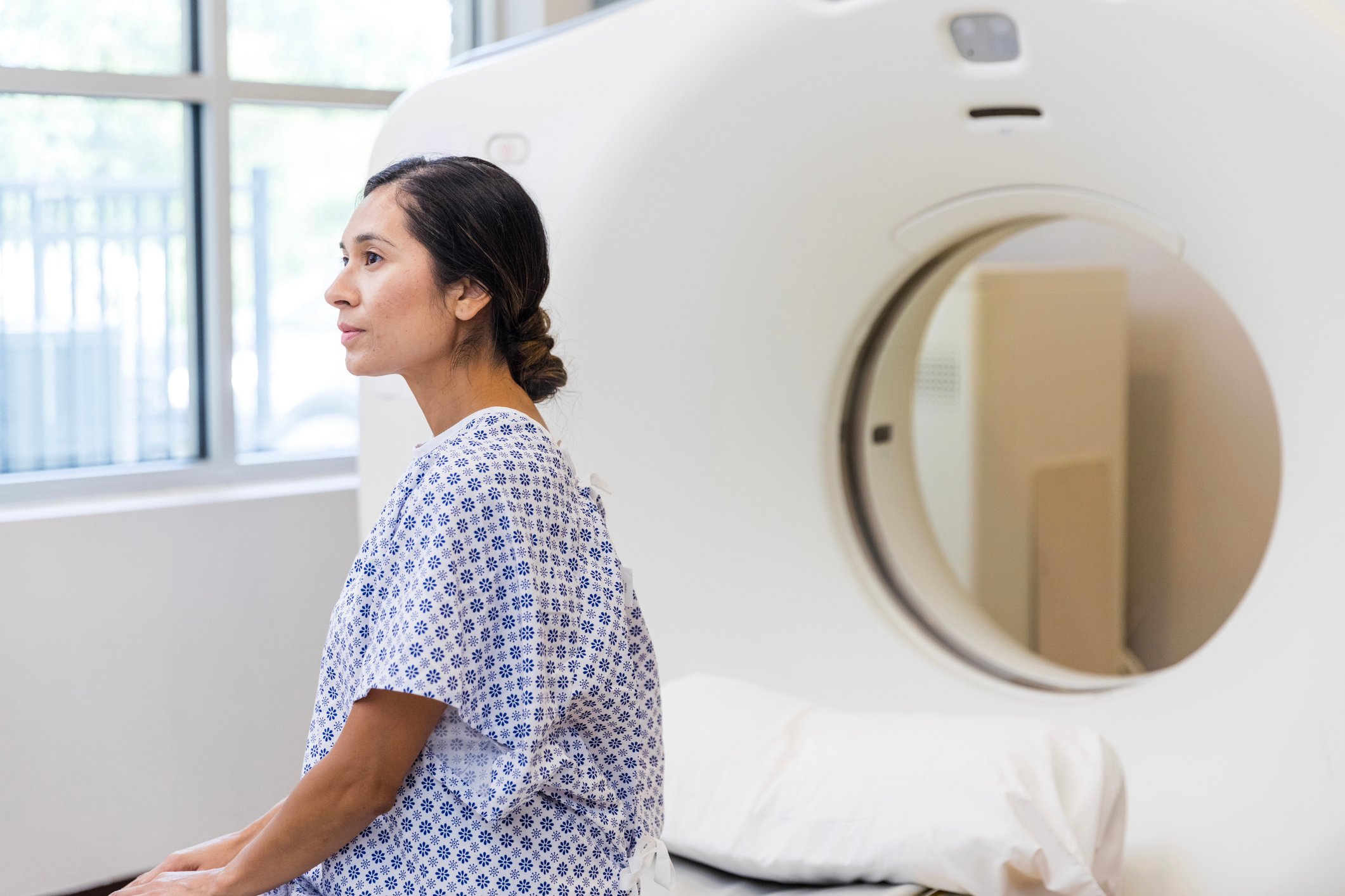
Your healthcare provider will tell you everything you need to know about CT scan preparation. Some general guidelines include:
- Plan to arrive early. Your provider will tell you when to come to your appointment
- Don’t eat for four hours before your CT scan
- Drink only clear liquids in the two hours leading up to your appointment
- Wear comfortable clothes and remove any metal jewelry or clothing. Your provider may give you a hospital gown to wear
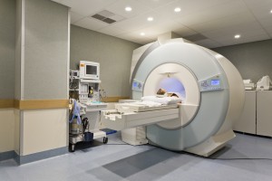
During the test, you will lie on your back on a table. If your test requires it, a healthcare provider may inject contrast dye intravenously. This dye can make you feel flushed or give you a metallic taste in your mouth. When the scan begins:
- The bed will slowly move in the doughnut-shaped scanner. At this point, you will need to stay as still as possible because movement can blur the images
- You may also be asked to hold your breath for a short period, usually fewer than 15 to 20 seconds
- The scanner takes pictures of the area your healthcare provider needs to see. Unlike an MRI scan, a CT scan is silent
- When the exam is over, the table moves back out of the scanner
You can have a CT scan in a hospital or an outpatient facility. CT scans usually take about an hour. However, with newer machines, scans can take only a few minutes. The whole process can take about 30 minutes.
After the exam, you can return to your normal routine. If you were given contrast dye, you may be asked to wait a short time before leaving to ensure that you feel okay after the exam. You might be told to drink lots of fluids to help your kidneys remove the dye from your body.
CT images are stored as electronic data files. They’re most often reviewed on a computer screen. A radiologist looks at the images and creates a report that is kept in your medical records. It usually takes about 24 to 48 hours to get the results of your CT scan. Your healthcare professional talks with you about the results.
To schedule an appointment with the Flushing Hospital Medical Center’s Radiology Department, call 718- 670-5458. To schedule an appointment for a CT scan or for more information about CT scans, please call 718-670-8851.
All content of this newsletter is intended for general information purposes only and is not intended or implied to be a substitute for professional medical advice, diagnosis or treatment. Please consult a medical professional before adopting any of the suggestions on this page. You must never disregard professional medical advice or delay seeking medical treatment based upon any content of this newsletter. PROMPTLY CONSULT YOUR PHYSICIAN OR CALL 911 IF YOU BELIEVE YOU HAVE A MEDICAL EMERGENCY.